 Add to Chrome
 Add to Firefox
 Add to Edge
Fernando Pérez-García
RAD-DINO: Exploring Scalable Medical Image Encoders Beyond Text Supervision
Jan 19, 2024
Language-supervised pre-training has proven to be a valuable method for extracting semantically meaningful features from images, serving as a foundational element in multimodal systems within the computer vision and medical imaging domains. However, resulting features are limited by the information contained within the text. This is particularly problematic in medical imaging, where radiologists' written findings focus on specific observations; a challenge compounded by the scarcity of paired imaging-text data due to concerns over leakage of personal health information. In this work, we fundamentally challenge the prevailing reliance on language supervision for learning general purpose biomedical imaging encoders. We introduce RAD-DINO, a biomedical image encoder pre-trained solely on unimodal biomedical imaging data that obtains similar or greater performance than state-of-the-art biomedical language supervised models on a diverse range of benchmarks. Specifically, the quality of learned representations is evaluated on standard imaging tasks (classification and semantic segmentation), and a vision-language alignment task (text report generation from images). To further demonstrate the drawback of language supervision, we show that features from RAD-DINO correlate with other medical records (e.g., sex or age) better than language-supervised models, which are generally not mentioned in radiology reports. Finally, we conduct a series of ablations determining the factors in RAD-DINO's performance; notably, we observe that RAD-DINO's downstream performance scales well with the quantity and diversity of training data, demonstrating that image-only supervision is a scalable approach for training a foundational biomedical image encoder.
RadEdit: stress-testing biomedical vision models via diffusion image editing
Dec 21, 2023Biomedical imaging datasets are often small and biased, meaning that real-world performance of predictive models can be substantially lower than expected from internal testing. This work proposes using generative image editing to simulate dataset shifts and diagnose failure modes of biomedical vision models; this can be used in advance of deployment to assess readiness, potentially reducing cost and patient harm. Existing editing methods can produce undesirable changes, with spurious correlations learned due to the co-occurrence of disease and treatment interventions, limiting practical applicability. To address this, we train a text-to-image diffusion model on multiple chest X-ray datasets and introduce a new editing method RadEdit that uses multiple masks, if present, to constrain changes and ensure consistency in the edited images. We consider three types of dataset shifts: acquisition shift, manifestation shift, and population shift, and demonstrate that our approach can diagnose failures and quantify model robustness without additional data collection, complementing more qualitative tools for explainable AI.
MAIRA-1: A specialised large multimodal model for radiology report generation
Nov 22, 2023We present a radiology-specific multimodal model for the task for generating radiological reports from chest X-rays (CXRs). Our work builds on the idea that large language model(s) can be equipped with multimodal capabilities through alignment with pre-trained vision encoders. On natural images, this has been shown to allow multimodal models to gain image understanding and description capabilities. Our proposed model (MAIRA-1) leverages a CXR-specific image encoder in conjunction with a fine-tuned large language model based on Vicuna-7B, and text-based data augmentation, to produce reports with state-of-the-art quality. In particular, MAIRA-1 significantly improves on the radiologist-aligned RadCliQ metric and across all lexical metrics considered. Manual review of model outputs demonstrates promising fluency and accuracy of generated reports while uncovering failure modes not captured by existing evaluation practices. More information and resources can be found on the project website: https://aka.ms/maira.
Exploring the Boundaries of GPT-4 in Radiology
Oct 23, 2023The recent success of general-domain large language models (LLMs) has significantly changed the natural language processing paradigm towards a unified foundation model across domains and applications. In this paper, we focus on assessing the performance of GPT-4, the most capable LLM so far, on the text-based applications for radiology reports, comparing against state-of-the-art (SOTA) radiology-specific models. Exploring various prompting strategies, we evaluated GPT-4 on a diverse range of common radiology tasks and we found GPT-4 either outperforms or is on par with current SOTA radiology models. With zero-shot prompting, GPT-4 already obtains substantial gains ($\approx$ 10% absolute improvement) over radiology models in temporal sentence similarity classification (accuracy) and natural language inference ($F_1$). For tasks that require learning dataset-specific style or schema (e.g. findings summarisation), GPT-4 improves with example-based prompting and matches supervised SOTA. Our extensive error analysis with a board-certified radiologist shows GPT-4 has a sufficient level of radiology knowledge with only occasional errors in complex context that require nuanced domain knowledge. For findings summarisation, GPT-4 outputs are found to be overall comparable with existing manually-written impressions.
Region-based Contrastive Pretraining for Medical Image Retrieval with Anatomic Query
May 09, 2023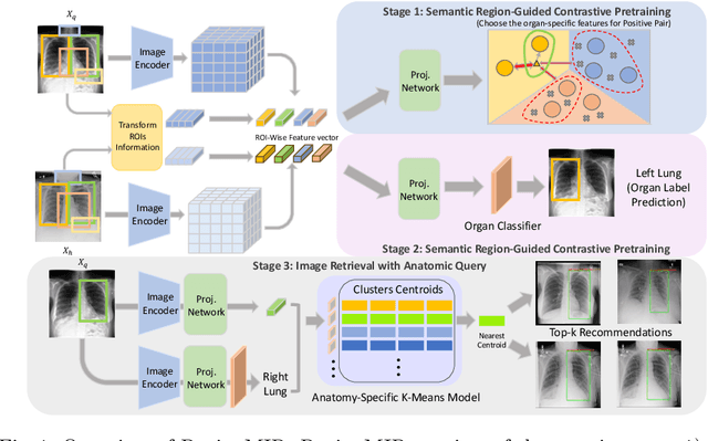
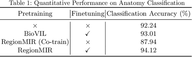
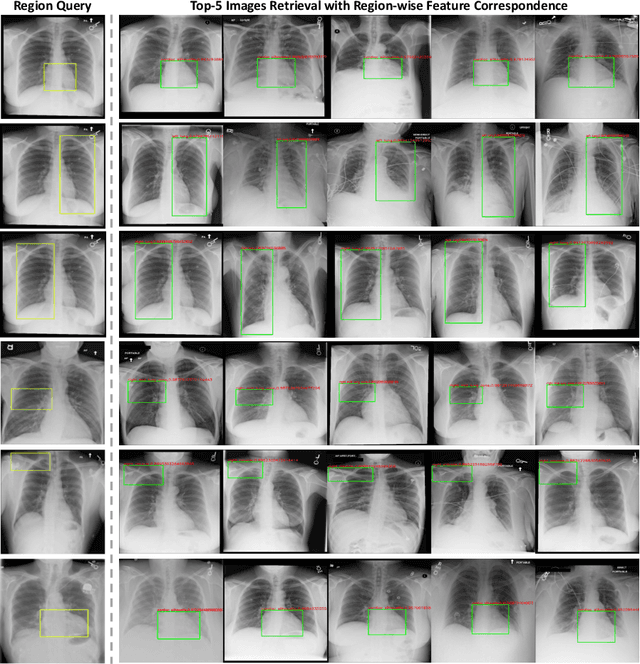
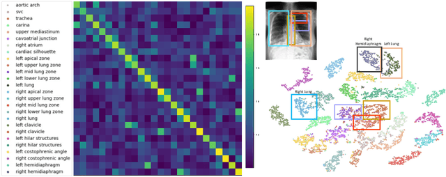
We introduce a novel Region-based contrastive pretraining for Medical Image Retrieval (RegionMIR) that demonstrates the feasibility of medical image retrieval with similar anatomical regions. RegionMIR addresses two major challenges for medical image retrieval i) standardization of clinically relevant searching criteria (e.g., anatomical, pathology-based), and ii) localization of anatomical area of interests that are semantically meaningful. In this work, we propose an ROI image retrieval image network that retrieves images with similar anatomy by extracting anatomical features (via bounding boxes) and evaluate similarity between pairwise anatomy-categorized features between the query and the database of images using contrastive learning. ROI queries are encoded using a contrastive-pretrained encoder that was fine-tuned for anatomy classification, which generates an anatomical-specific latent space for region-correlated image retrieval. During retrieval, we compare the anatomically encoded query to find similar features within a feature database generated from training samples, and retrieve images with similar regions from training samples. We evaluate our approach on both anatomy classification and image retrieval tasks using the Chest ImaGenome Dataset. Our proposed strategy yields an improvement over state-of-the-art pretraining and co-training strategies, from 92.24 to 94.12 (2.03%) classification accuracy in anatomies. We qualitatively evaluate the image retrieval performance demonstrating generalizability across multiple anatomies with different morphology.
Compositional Zero-Shot Domain Transfer with Text-to-Text Models
Mar 23, 2023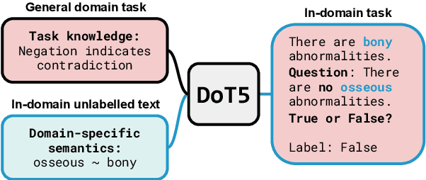
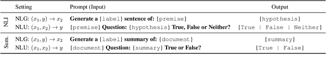
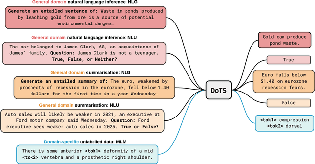
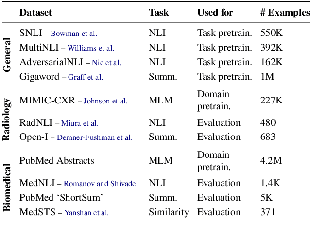
Label scarcity is a bottleneck for improving task performance in specialised domains. We propose a novel compositional transfer learning framework (DoT5 - domain compositional zero-shot T5) for zero-shot domain transfer. Without access to in-domain labels, DoT5 jointly learns domain knowledge (from MLM of unlabelled in-domain free text) and task knowledge (from task training on more readily available general-domain data) in a multi-task manner. To improve the transferability of task training, we design a strategy named NLGU: we simultaneously train NLG for in-domain label-to-data generation which enables data augmentation for self-finetuning and NLU for label prediction. We evaluate DoT5 on the biomedical domain and the resource-lean subdomain of radiology, focusing on NLI, text summarisation and embedding learning. DoT5 demonstrates the effectiveness of compositional transfer learning through multi-task learning. In particular, DoT5 outperforms the current SOTA in zero-shot transfer by over 7 absolute points in accuracy on RadNLI. We validate DoT5 with ablations and a case study demonstrating its ability to solve challenging NLI examples requiring in-domain expertise.
MONAI Label: A framework for AI-assisted Interactive Labeling of 3D Medical Images
Mar 23, 2022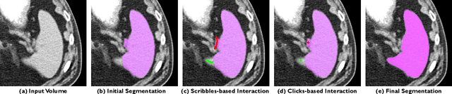
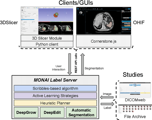
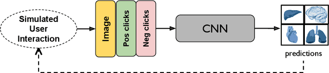
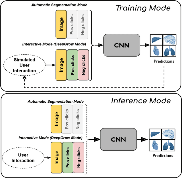
The lack of annotated datasets is a major challenge in training new task-specific supervised AI algorithms as manual annotation is expensive and time-consuming. To address this problem, we present MONAI Label, a free and open-source platform that facilitates the development of AI-based applications that aim at reducing the time required to annotate 3D medical image datasets. Through MONAI Label researchers can develop annotation applications focusing on their domain of expertise. It allows researchers to readily deploy their apps as services, which can be made available to clinicians via their preferred user-interface. Currently, MONAI Label readily supports locally installed (3DSlicer) and web-based (OHIF) frontends, and offers two Active learning strategies to facilitate and speed up the training of segmentation algorithms. MONAI Label allows researchers to make incremental improvements to their labeling apps by making them available to other researchers and clinicians alike. Lastly, MONAI Label provides sample labeling apps, namely DeepEdit and DeepGrow, demonstrating dramatically reduced annotation times.
Transfer Learning of Deep Spatiotemporal Networks to Model Arbitrarily Long Videos of Seizures
Jun 22, 2021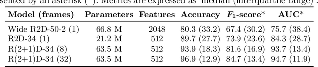
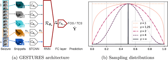
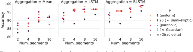
Detailed analysis of seizure semiology, the symptoms and signs which occur during a seizure, is critical for management of epilepsy patients. Inter-rater reliability using qualitative visual analysis is often poor for semiological features. Therefore, automatic and quantitative analysis of video-recorded seizures is needed for objective assessment. We present GESTURES, a novel architecture combining convolutional neural networks (CNNs) and recurrent neural networks (RNNs) to learn deep representations of arbitrarily long videos of epileptic seizures. We use a spatiotemporal CNN (STCNN) pre-trained on large human action recognition (HAR) datasets to extract features from short snippets (approx. 0.5 s) sampled from seizure videos. We then train an RNN to learn seizure-level representations from the sequence of features. We curated a dataset of seizure videos from 68 patients and evaluated GESTURES on its ability to classify seizures into focal onset seizures (FOSs) (N = 106) vs. focal to bilateral tonic-clonic seizures (TCSs) (N = 77), obtaining an accuracy of 98.9% using bidirectional long short-term memory (BLSTM) units. We demonstrate that an STCNN trained on a HAR dataset can be used in combination with an RNN to accurately represent arbitrarily long videos of seizures. GESTURES can provide accurate seizure classification by modeling sequences of semiologies.
A self-supervised learning strategy for postoperative brain cavity segmentation simulating resections
May 24, 2021
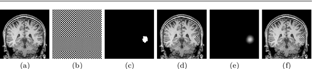
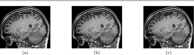

Accurate segmentation of brain resection cavities (RCs) aids in postoperative analysis and determining follow-up treatment. Convolutional neural networks (CNNs) are the state-of-the-art image segmentation technique, but require large annotated datasets for training. Annotation of 3D medical images is time-consuming, requires highly-trained raters, and may suffer from high inter-rater variability. Self-supervised learning strategies can leverage unlabeled data for training. We developed an algorithm to simulate resections from preoperative magnetic resonance images (MRIs). We performed self-supervised training of a 3D CNN for RC segmentation using our simulation method. We curated EPISURG, a dataset comprising 430 postoperative and 268 preoperative MRIs from 430 refractory epilepsy patients who underwent resective neurosurgery. We fine-tuned our model on three small annotated datasets from different institutions and on the annotated images in EPISURG, comprising 20, 33, 19 and 133 subjects. The model trained on data with simulated resections obtained median (interquartile range) Dice score coefficients (DSCs) of 81.7 (16.4), 82.4 (36.4), 74.9 (24.2) and 80.5 (18.7) for each of the four datasets. After fine-tuning, DSCs were 89.2 (13.3), 84.1 (19.8), 80.2 (20.1) and 85.2 (10.8). For comparison, inter-rater agreement between human annotators from our previous study was 84.0 (9.9). We present a self-supervised learning strategy for 3D CNNs using simulated RCs to accurately segment real RCs on postoperative MRI. Our method generalizes well to data from different institutions, pathologies and modalities. Source code, segmentation models and the EPISURG dataset are available at https://github.com/fepegar/ressegijcars .
Simulation of Brain Resection for Cavity Segmentation Using Self-Supervised and Semi-Supervised Learning
Jun 28, 2020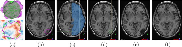
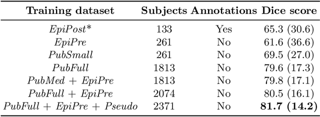
Resective surgery may be curative for drug-resistant focal epilepsy, but only 40% to 70% of patients achieve seizure freedom after surgery. Retrospective quantitative analysis could elucidate patterns in resected structures and patient outcomes to improve resective surgery. However, the resection cavity must first be segmented on the postoperative MR image. Convolutional neural networks (CNNs) are the state-of-the-art image segmentation technique, but require large amounts of annotated data for training. Annotation of medical images is a time-consuming process requiring highly-trained raters, and often suffering from high inter-rater variability. Self-supervised learning can be used to generate training instances from unlabeled data. We developed an algorithm to simulate resections on preoperative MR images. We curated a new dataset, EPISURG, comprising 431 postoperative and 269 preoperative MR images from 431 patients who underwent resective surgery. In addition to EPISURG, we used three public datasets comprising 1813 preoperative MR images for training. We trained a 3D CNN on artificially resected images created on the fly during training, using images from 1) EPISURG, 2) public datasets and 3) both. To evaluate trained models, we calculate Dice score (DSC) between model segmentations and 200 manual annotations performed by three human raters. The model trained on data with manual annotations obtained a median (interquartile range) DSC of 65.3 (30.6). The DSC of our best-performing model, trained with no manual annotations, is 81.7 (14.2). For comparison, inter-rater agreement between human annotators was 84.0 (9.9). We demonstrate a training method for CNNs using simulated resection cavities that can accurately segment real resection cavities, without manual annotations.