 Add to Chrome
 Add to Firefox
 Add to Edge
Guanghua Xiao
Discovering Clinically Meaningful Shape Features for the Analysis of Tumor Pathology Images
Dec 09, 2020
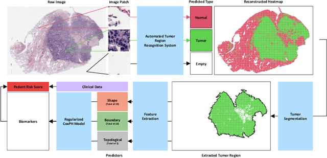
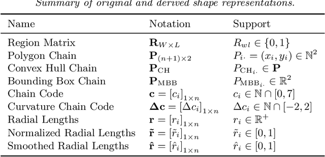
With the advanced imaging technology, digital pathology imaging of tumor tissue slides is becoming a routine clinical procedure for cancer diagnosis. This process produces massive imaging data that capture histological details in high resolution. Recent developments in deep-learning methods have enabled us to automatically detect and characterize the tumor regions in pathology images at large scale. From each identified tumor region, we extracted 30 well-defined descriptors that quantify its shape, geometry, and topology. We demonstrated how those descriptor features were associated with patient survival outcome in lung adenocarcinoma patients from the National Lung Screening Trial (n=143). Besides, a descriptor-based prognostic model was developed and validated in an independent patient cohort from The Cancer Genome Atlas Program program (n=318). This study proposes new insights into the relationship between tumor shape, geometrical, and topological features and patient prognosis. We provide software in the form of R code on GitHub: https://github.com/estfernandez/Slide_Image_Segmentation_and_Extraction.
Predicting survival outcomes using topological features of tumor pathology images
Dec 07, 2020
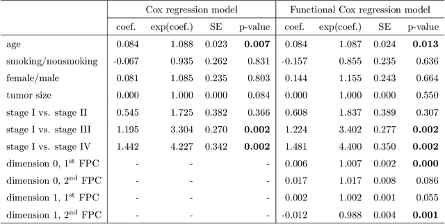
Tumor shape and size have been used as important markers for cancer diagnosis and treatment. Recent developments in medical imaging technology enable more detailed segmentation of tumor regions in high resolution. This paper proposes a topological feature to characterize tumor progression from digital pathology images and examine its effect on the time-to-event data. We develop distance transform for pathology images and show that a topological summary statistic computed by persistent homology quantifies tumor shape, size, distribution, and connectivity. The topological features are represented in functional space and used as functional predictors in a functional Cox regression model. A case study is conducted using non-small cell lung cancer pathology images. The results show that the topological features predict survival prognosis after adjusting for age, sex, smoking status, stage, and size of tumors. Also, the topological features with non-zero effects correspond to the shapes that are known to be related to tumor progression. Our study provides a new perspective for understanding tumor shape and patient prognosis.
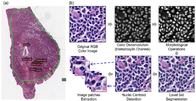
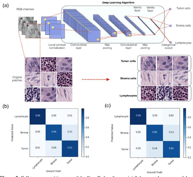
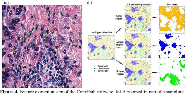
ConvPath: A Software Tool for Lung Adenocarcinoma Digital Pathological Image Analysis Aided by Convolutional Neural Network
Sep 20, 2018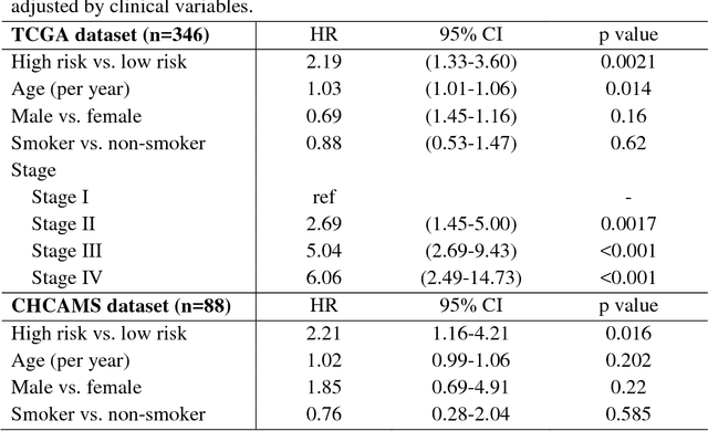
The spatial distributions of different types of cells could reveal a cancer cell growth pattern, its relationships with the tumor microenvironment and the immune response of the body, all of which represent key hallmarks of cancer. However, manually recognizing and localizing all the cells in pathology slides are almost impossible. In this study, we developed an automated cell type classification pipeline, ConvPath, which includes nuclei segmentation, convolutional neural network-based tumor, stromal and lymphocytes classification, and extraction of tumor microenvironment related features for lung cancer pathology images. The overall classification accuracy is 92.9% and 90.1% in training and independent testing datasets, respectively. By identifying cells and classifying cell types, this pipeline can convert a pathology image into a spatial map of tumor, stromal and lymphocyte cells. From this spatial map, we can extracted features that characterize the tumor micro-environment. Based on these features, we developed an image feature-based prognostic model and validated the model in two independent cohorts. The predicted risk group serves as an independent prognostic factor, after adjusting for clinical variables that include age, gender, smoking status, and stage.