 Add to Chrome
 Add to Firefox
 Add to Edge
Shaoting Zhang
FPL+: Filtered Pseudo Label-based Unsupervised Cross-Modality Adaptation for 3D Medical Image Segmentation
Apr 07, 2024
Adapting a medical image segmentation model to a new domain is important for improving its cross-domain transferability, and due to the expensive annotation process, Unsupervised Domain Adaptation (UDA) is appealing where only unlabeled images are needed for the adaptation. Existing UDA methods are mainly based on image or feature alignment with adversarial training for regularization, and they are limited by insufficient supervision in the target domain. In this paper, we propose an enhanced Filtered Pseudo Label (FPL+)-based UDA method for 3D medical image segmentation. It first uses cross-domain data augmentation to translate labeled images in the source domain to a dual-domain training set consisting of a pseudo source-domain set and a pseudo target-domain set. To leverage the dual-domain augmented images to train a pseudo label generator, domain-specific batch normalization layers are used to deal with the domain shift while learning the domain-invariant structure features, generating high-quality pseudo labels for target-domain images. We then combine labeled source-domain images and target-domain images with pseudo labels to train a final segmentor, where image-level weighting based on uncertainty estimation and pixel-level weighting based on dual-domain consensus are proposed to mitigate the adverse effect of noisy pseudo labels. Experiments on three public multi-modal datasets for Vestibular Schwannoma, brain tumor and whole heart segmentation show that our method surpassed ten state-of-the-art UDA methods, and it even achieved better results than fully supervised learning in the target domain in some cases.
PathoTune: Adapting Visual Foundation Model to Pathological Specialists
Mar 25, 2024As natural image understanding moves towards the pretrain-finetune era, research in pathology imaging is concurrently evolving. Despite the predominant focus on pretraining pathological foundation models, how to adapt foundation models to downstream tasks is little explored. For downstream adaptation, we propose the existence of two domain gaps, i.e., the Foundation-Task Gap and the Task-Instance Gap. To mitigate these gaps, we introduce PathoTune, a framework designed to efficiently adapt pathological or even visual foundation models to pathology-specific tasks via multi-modal prompt tuning. The proposed framework leverages Task-specific Visual Prompts and Task-specific Textual Prompts to identify task-relevant features, along with Instance-specific Visual Prompts for encoding single pathological image features. Results across multiple datasets at both patch-level and WSI-level demonstrate its superior performance over single-modality prompt tuning approaches. Significantly, PathoTune facilitates the direct adaptation of natural visual foundation models to pathological tasks, drastically outperforming pathological foundation models with simple linear probing. The code will be available upon acceptance.
VLM-CPL: Consensus Pseudo Labels from Vision-Language Models for Human Annotation-Free Pathological Image Classification
Mar 23, 2024Despite that deep learning methods have achieved remarkable performance in pathology image classification, they heavily rely on labeled data, demanding extensive human annotation efforts. In this study, we present a novel human annotation-free method for pathology image classification by leveraging pre-trained Vision-Language Models (VLMs). Without human annotation, pseudo labels of the training set are obtained by utilizing the zero-shot inference capabilities of VLM, which may contain a lot of noise due to the domain shift between the pre-training data and the target dataset. To address this issue, we introduce VLM-CPL, a novel approach based on consensus pseudo labels that integrates two noisy label filtering techniques with a semi-supervised learning strategy. Specifically, we first obtain prompt-based pseudo labels with uncertainty estimation by zero-shot inference with the VLM using multiple augmented views of an input. Then, by leveraging the feature representation ability of VLM, we obtain feature-based pseudo labels via sample clustering in the feature space. Prompt-feature consensus is introduced to select reliable samples based on the consensus between the two types of pseudo labels. By rejecting low-quality pseudo labels, we further propose High-confidence Cross Supervision (HCS) to learn from samples with reliable pseudo labels and the remaining unlabeled samples. Experimental results showed that our method obtained an accuracy of 87.1% and 95.1% on the HPH and LC25K datasets, respectively, and it largely outperformed existing zero-shot classification and noisy label learning methods. The code is available at https://github.com/lanfz2000/VLM-CPL.
Modality-Aware and Shift Mixer for Multi-modal Brain Tumor Segmentation
Mar 04, 2024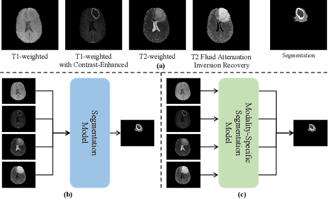
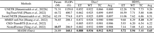
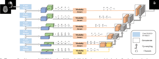
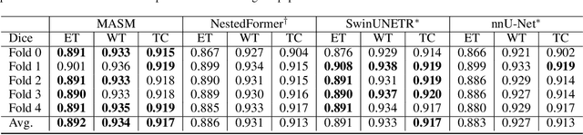
Combining images from multi-modalities is beneficial to explore various information in computer vision, especially in the medical domain. As an essential part of clinical diagnosis, multi-modal brain tumor segmentation aims to delineate the malignant entity involving multiple modalities. Although existing methods have shown remarkable performance in the task, the information exchange for cross-scale and high-level representations fusion in spatial and modality are limited in these methods. In this paper, we present a novel Modality Aware and Shift Mixer that integrates intra-modality and inter-modality dependencies of multi-modal images for effective and robust brain tumor segmentation. Specifically, we introduce a Modality-Aware module according to neuroimaging studies for modeling the specific modality pair relationships at low levels, and a Modality-Shift module with specific mosaic patterns is developed to explore the complex relationships across modalities at high levels via the self-attention. Experimentally, we outperform previous state-of-the-art approaches on the public Brain Tumor Segmentation (BraTS 2021 segmentation) dataset. Further qualitative experiments demonstrate the efficacy and robustness of MASM.
OpenMEDLab: An Open-source Platform for Multi-modality Foundation Models in Medicine
Mar 04, 2024The emerging trend of advancing generalist artificial intelligence, such as GPTv4 and Gemini, has reshaped the landscape of research (academia and industry) in machine learning and many other research areas. However, domain-specific applications of such foundation models (e.g., in medicine) remain untouched or often at their very early stages. It will require an individual set of transfer learning and model adaptation techniques by further expanding and injecting these models with domain knowledge and data. The development of such technologies could be largely accelerated if the bundle of data, algorithms, and pre-trained foundation models were gathered together and open-sourced in an organized manner. In this work, we present OpenMEDLab, an open-source platform for multi-modality foundation models. It encapsulates not only solutions of pioneering attempts in prompting and fine-tuning large language and vision models for frontline clinical and bioinformatic applications but also building domain-specific foundation models with large-scale multi-modal medical data. Importantly, it opens access to a group of pre-trained foundation models for various medical image modalities, clinical text, protein engineering, etc. Inspiring and competitive results are also demonstrated for each collected approach and model in a variety of benchmarks for downstream tasks. We welcome researchers in the field of medical artificial intelligence to continuously contribute cutting-edge methods and models to OpenMEDLab, which can be accessed via https://github.com/openmedlab.
Swin-UMamba: Mamba-based UNet with ImageNet-based pretraining
Feb 05, 2024Accurate medical image segmentation demands the integration of multi-scale information, spanning from local features to global dependencies. However, it is challenging for existing methods to model long-range global information, where convolutional neural networks (CNNs) are constrained by their local receptive fields, and vision transformers (ViTs) suffer from high quadratic complexity of their attention mechanism. Recently, Mamba-based models have gained great attention for their impressive ability in long sequence modeling. Several studies have demonstrated that these models can outperform popular vision models in various tasks, offering higher accuracy, lower memory consumption, and less computational burden. However, existing Mamba-based models are mostly trained from scratch and do not explore the power of pretraining, which has been proven to be quite effective for data-efficient medical image analysis. This paper introduces a novel Mamba-based model, Swin-UMamba, designed specifically for medical image segmentation tasks, leveraging the advantages of ImageNet-based pretraining. Our experimental results reveal the vital role of ImageNet-based training in enhancing the performance of Mamba-based models. Swin-UMamba demonstrates superior performance with a large margin compared to CNNs, ViTs, and latest Mamba-based models. Notably, on AbdomenMRI, Encoscopy, and Microscopy datasets, Swin-UMamba outperforms its closest counterpart U-Mamba by an average score of 3.58%. The code and models of Swin-UMamba are publicly available at: https://github.com/JiarunLiu/Swin-UMamba
Evaluating and Enhancing Large Language Models Performance in Domain-specific Medicine: Osteoarthritis Management with DocOA
Jan 20, 2024The efficacy of large language models (LLMs) in domain-specific medicine, particularly for managing complex diseases such as osteoarthritis (OA), remains largely unexplored. This study focused on evaluating and enhancing the clinical capabilities of LLMs in specific domains, using osteoarthritis (OA) management as a case study. A domain specific benchmark framework was developed, which evaluate LLMs across a spectrum from domain-specific knowledge to clinical applications in real-world clinical scenarios. DocOA, a specialized LLM tailored for OA management that integrates retrieval-augmented generation (RAG) and instruction prompts, was developed. The study compared the performance of GPT-3.5, GPT-4, and a specialized assistant, DocOA, using objective and human evaluations. Results showed that general LLMs like GPT-3.5 and GPT-4 were less effective in the specialized domain of OA management, particularly in providing personalized treatment recommendations. However, DocOA showed significant improvements. This study introduces a novel benchmark framework which assesses the domain-specific abilities of LLMs in multiple aspects, highlights the limitations of generalized LLMs in clinical contexts, and demonstrates the potential of tailored approaches for developing domain-specific medical LLMs.
Data-Centric Foundation Models in Computational Healthcare: A Survey
Jan 04, 2024The advent of foundation models (FMs) as an emerging suite of AI techniques has struck a wave of opportunities in computational healthcare. The interactive nature of these models, guided by pre-training data and human instructions, has ignited a data-centric AI paradigm that emphasizes better data characterization, quality, and scale. In healthcare AI, obtaining and processing high-quality clinical data records has been a longstanding challenge, ranging from data quantity, annotation, patient privacy, and ethics. In this survey, we investigate a wide range of data-centric approaches in the FM era (from model pre-training to inference) towards improving the healthcare workflow. We discuss key perspectives in AI security, assessment, and alignment with human values. Finally, we offer a promising outlook of FM-based analytics to enhance the performance of patient outcome and clinical workflow in the evolving landscape of healthcare and medicine. We provide an up-to-date list of healthcare-related foundation models and datasets at https://github.com/Yunkun-Zhang/Data-Centric-FM-Healthcare .
SegRap2023: A Benchmark of Organs-at-Risk and Gross Tumor Volume Segmentation for Radiotherapy Planning of Nasopharyngeal Carcinoma
Dec 15, 2023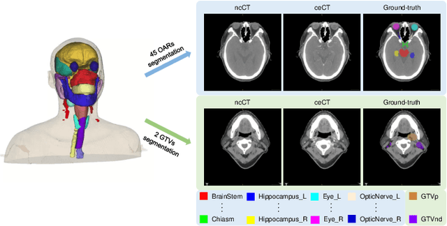
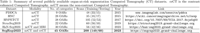
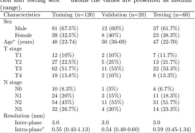
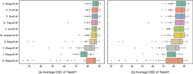
Radiation therapy is a primary and effective NasoPharyngeal Carcinoma (NPC) treatment strategy. The precise delineation of Gross Tumor Volumes (GTVs) and Organs-At-Risk (OARs) is crucial in radiation treatment, directly impacting patient prognosis. Previously, the delineation of GTVs and OARs was performed by experienced radiation oncologists. Recently, deep learning has achieved promising results in many medical image segmentation tasks. However, for NPC OARs and GTVs segmentation, few public datasets are available for model development and evaluation. To alleviate this problem, the SegRap2023 challenge was organized in conjunction with MICCAI2023 and presented a large-scale benchmark for OAR and GTV segmentation with 400 Computed Tomography (CT) scans from 200 NPC patients, each with a pair of pre-aligned non-contrast and contrast-enhanced CT scans. The challenge's goal was to segment 45 OARs and 2 GTVs from the paired CT scans. In this paper, we detail the challenge and analyze the solutions of all participants. The average Dice similarity coefficient scores for all submissions ranged from 76.68\% to 86.70\%, and 70.42\% to 73.44\% for OARs and GTVs, respectively. We conclude that the segmentation of large-size OARs is well-addressed, and more efforts are needed for GTVs and small-size or thin-structure OARs. The benchmark will remain publicly available here: https://segrap2023.grand-challenge.org
ZePT: Zero-Shot Pan-Tumor Segmentation via Query-Disentangling and Self-Prompting
Dec 07, 2023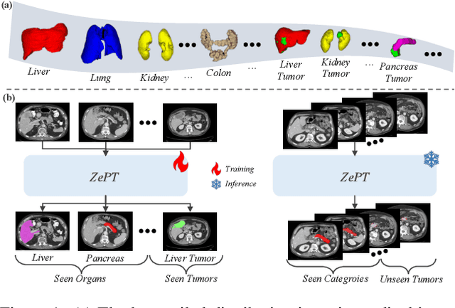
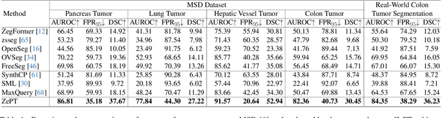
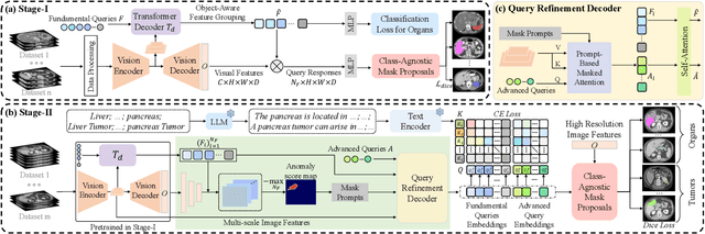
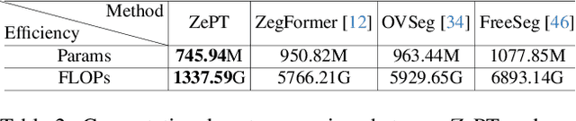
The long-tailed distribution problem in medical image analysis reflects a high prevalence of common conditions and a low prevalence of rare ones, which poses a significant challenge in developing a unified model capable of identifying rare or novel tumor categories not encountered during training. In this paper, we propose a new zero-shot pan-tumor segmentation framework (ZePT) based on query-disentangling and self-prompting to segment unseen tumor categories beyond the training set. ZePT disentangles the object queries into two subsets and trains them in two stages. Initially, it learns a set of fundamental queries for organ segmentation through an object-aware feature grouping strategy, which gathers organ-level visual features. Subsequently, it refines the other set of advanced queries that focus on the auto-generated visual prompts for unseen tumor segmentation. Moreover, we introduce query-knowledge alignment at the feature level to enhance each query's discriminative representation and generalizability. Extensive experiments on various tumor segmentation tasks demonstrate the performance superiority of ZePT, which surpasses the previous counterparts and evidence the promising ability for zero-shot tumor segmentation in real-world settings. Codes will be made publicly available.